
 Powered by Bioz
PART/ 85108075013
pco.edge 4.2 bi UV USB sCMOS Camera
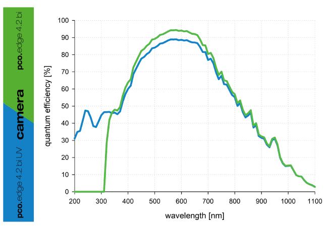
The pco.edge 4.2 bi UV is built on the trusted pco.edge series of scientific CMOS cameras with modern back-illuminated sensor technology. It offers a 2048 x 2048 pixel high resolution and 6.5 x 6.5 μm² pixel size for high quality imaging. Featuring a specialized input window, this UV sensitive sCMOS camera can achieve high quantum efficiency in the ultraviolet wavelength range.
The pco.edge 4.2 bi UV USB sCMOS camera offers an adjustable cooling system with the option to use air or water to cool the sensor down to -25 °C thus, reducing the dark current to 0.2 e-/pixel/s. It also incorporates a powerful USB3.1 interface complementing its high performance.
| Interface | USB 3.1 Gen 1 |
| Sensor technology | back-illuminated sCMOS |
| Color type | monochrome |
| Resolution | 2048 x 2048 pixels |
| Sensor diagonal | 18.8 mm |
| Pixel size | 6.5 x 6.5 μm |
| Maximum frame rate @ full resolution | 40 fps |
| Maximum pixel rate | 184 MPixel/s |
| Peak QE | 89 % @ 580 nm |
| Typical read noise1 | 1.0 e- |
| Dark current @ sensor temperature | < 0.2 @ -25 °C e-/pixel/s |
| Maximum dynamic range | 26,667 : 1 |
| Shutter type | Rolling Shutter, Global Reset |
| Sensor cooling2 | air & water |
| Additional options | lightsheet scanning mode3, |
| Dimensions (H x W x L) | 85 x 80 x 109mm |
- Brightfield microscopy microscopy
- Fluorescence microscopy
- Digital pathology
- Single molecule localization microscopy
- Lightsheet fluorescence microscopy (LSFM)
- Calcium imaging
- FRET
- FRAP
- Structured illumination microscopy (SIM)
- Highspeed bright field ratio imaging
- High throughput screening
- High content screening
- Biochip reading
- TIRF microscopy
- Spinning disk confocal microscopy
- 3D metrology
- Ophthalmology
- Photovoltaic inspection
- Industrial quality inspection
- Lucky astronomy
- Bio luminescence
- Chemo luminescence
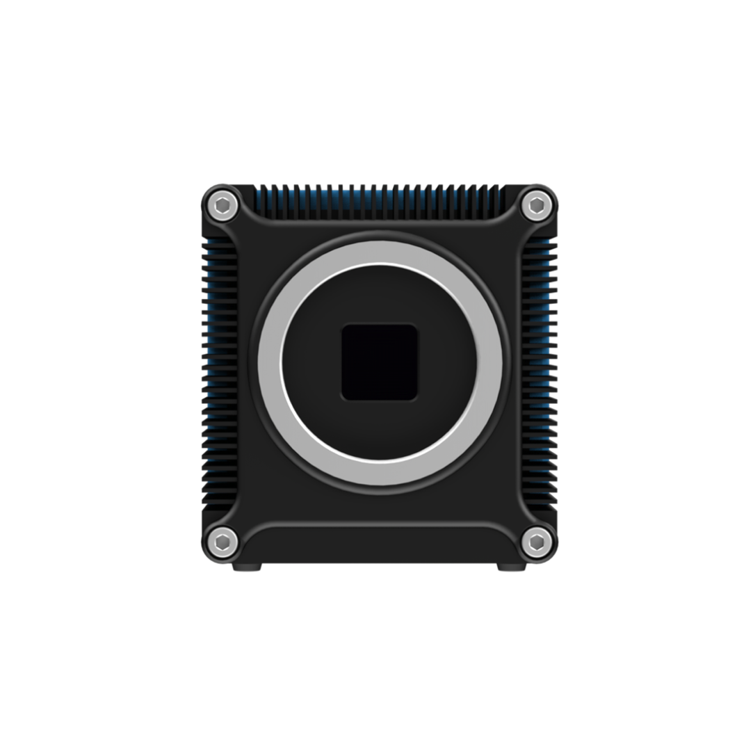
Front View
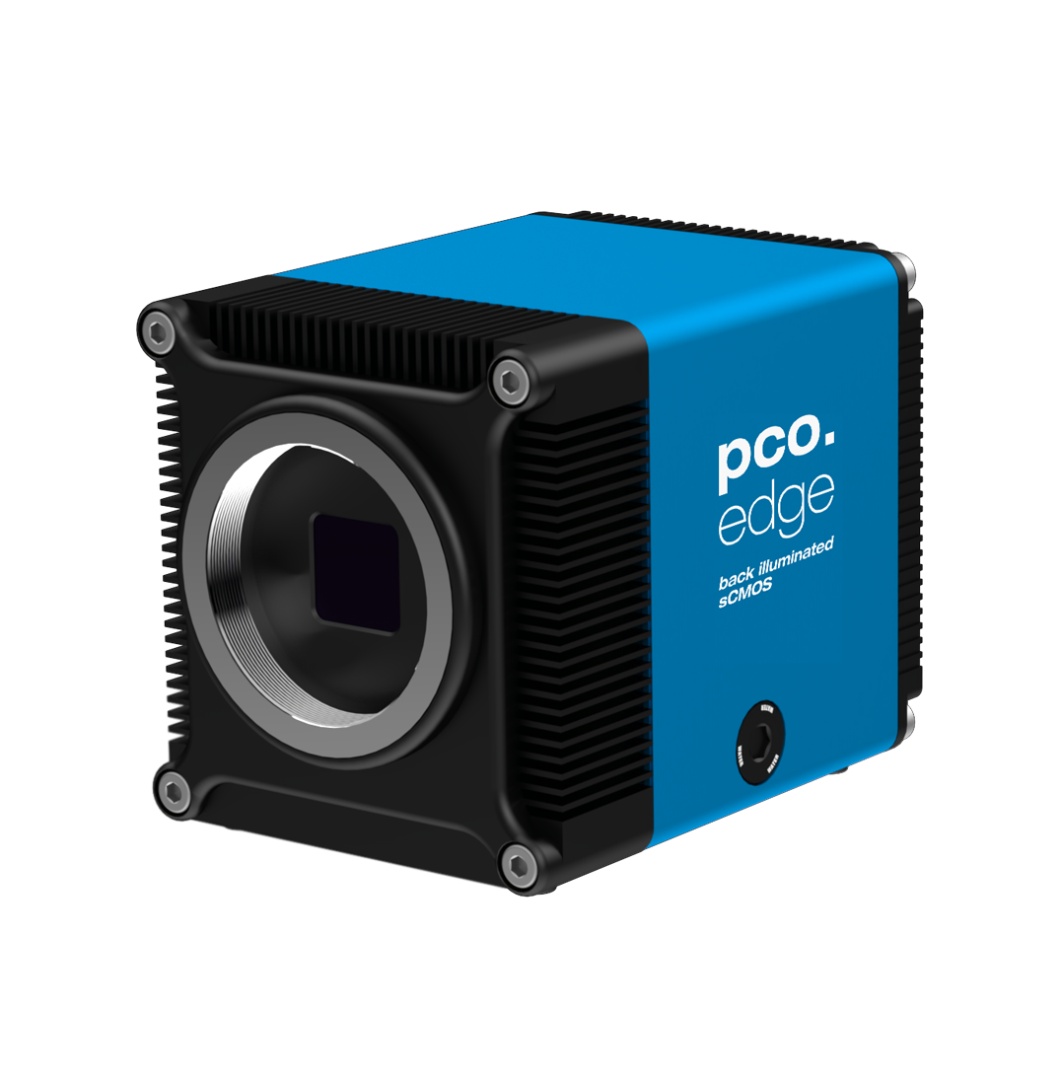
Front View 45 Degree
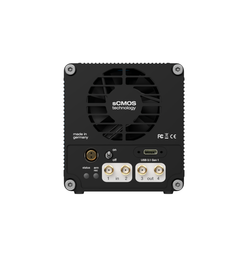
Rear View
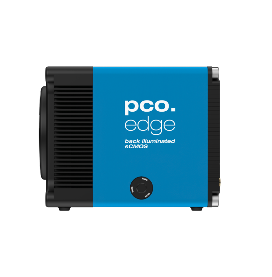
Side View
| Interface | USB 3.1 Gen 1 |
| Sensor technology | back-illuminated sCMOS |
| Color type | monochrome |
| Resolution | 2048 x 2048 pixels |
| Sensor diagonal | 18.8 mm |
| Pixel size | 6.5 x 6.5 μm |
| Maximum frame rate @ full resolution | 40 fps |
| Maximum pixel rate | 184 MPixel/s |
| Peak QE | 89 % @ 580 nm |
| Typical read noise1 | 1.0 e- |
| Dark current @ sensor temperature | < 0.2 @ -25 °C e-/pixel/s |
| Maximum dynamic range | 26,667 : 1 |
| Shutter type | Rolling Shutter, Global Reset |
| Sensor cooling2 | air & water |
| Additional options | lightsheet scanning mode3, |
| Dimensions (H x W x L) | 85 x 80 x 109mm |
- Brightfield microscopy microscopy
- Fluorescence microscopy
- Digital pathology
- Single molecule localization microscopy
- Lightsheet fluorescence microscopy (LSFM)
- Calcium imaging
- FRET
- FRAP
- Structured illumination microscopy (SIM)
- Highspeed bright field ratio imaging
- High throughput screening
- High content screening
- Biochip reading
- TIRF microscopy
- Spinning disk confocal microscopy
- 3D metrology
- Ophthalmology
- Photovoltaic inspection
- Industrial quality inspection
- Lucky astronomy
- Bio luminescence
- Chemo luminescence
Front View
Front View 45 Degree
Rear View
Side View
Subscribe Now!
Floating Web Forms block
Join the PCO Circular newsletter for early access to insights on our high‑performance imaging cameras. Receive the latest application notes, expert webinars, technical articles, educational videos, product innovations, and industry events designed for scientific, microscopy, research, and industrial imaging applications.