
 Powered by Bioz
pco.edge 4.2 CLHS sCMOS Camera
The pco.edge 4.2 is a high resolution, cooled sCMOS camera that operates with streaming via a 10G FOL (Camera Link HS) to allow direct processing of large data transfers through its high-performance data interface making it easy to securely bridge distances of up to 10 km. The pco.edge 4.2 is equipped with a low noise 16-bit scientific CMOS sensor to ensure crisp images and precise measurements. The pco.edge 4.2 sCMOS camera also comes with an upgrade option to include a water cooling system.
| Interface | CLHS FOL |
| Sensor technology | sCMOS |
| Color type | monochrome |
| Resolution | 2048 x 2048 pixels |
| Sensor diagonal | 18.8 mm |
| Pixel size | 6.5 x 6.5 μm |
| Maximum frame rate @ full resolution | 100 fps |
| Maximum pixel rate | 548 MPixel/s |
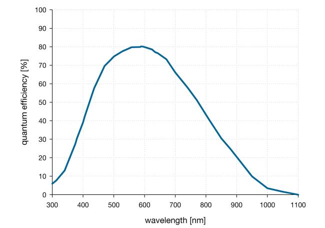
| Peak QE | 82 % @ 580 nm |
| Typical read noise1 | 0.8 e- |
| Dark current @ sensor temperature | < 0.6 @ 7 °C e-/pixel/s |
| Maximum dynamic range | 37,500 : 1 |
| Shutter type | Rolling Shutter |
| Sensor cooling2 | air, optional: water |
| Additional options | lens control |
| Dimensions (H x W x L) | 76 x 70 x 122 mm |
- Bright-field microscopy
- Fluorescence microscopy
- Digital pathology
- Single molecule localization microscopy (SMLM) – PALM, STORM, dSTORM, GSDIM
- Lightsheet fluorescence microscopy (LSFM)
- Structured illumination microscopy (SIM)
- Calcium imaging
- Förster resonance energy transfer (FRET)
- Fluorescence recovery after photobleaching (FRAP)
- High-speed bright-field ratio imaging
- High throughput screening | high content screening
- Biochip reading
- Total internal reflection microscopy (TITF)
- Spinning disk confocal microscopy
- 3D metrology
- Ophthalmology
- Photovoltaic inspection
- Industrial quality inspection
- Wafer inspection
- Image intensifier imaging
- Lucky astronomy
- Particle tracking velocimetry (PTV)
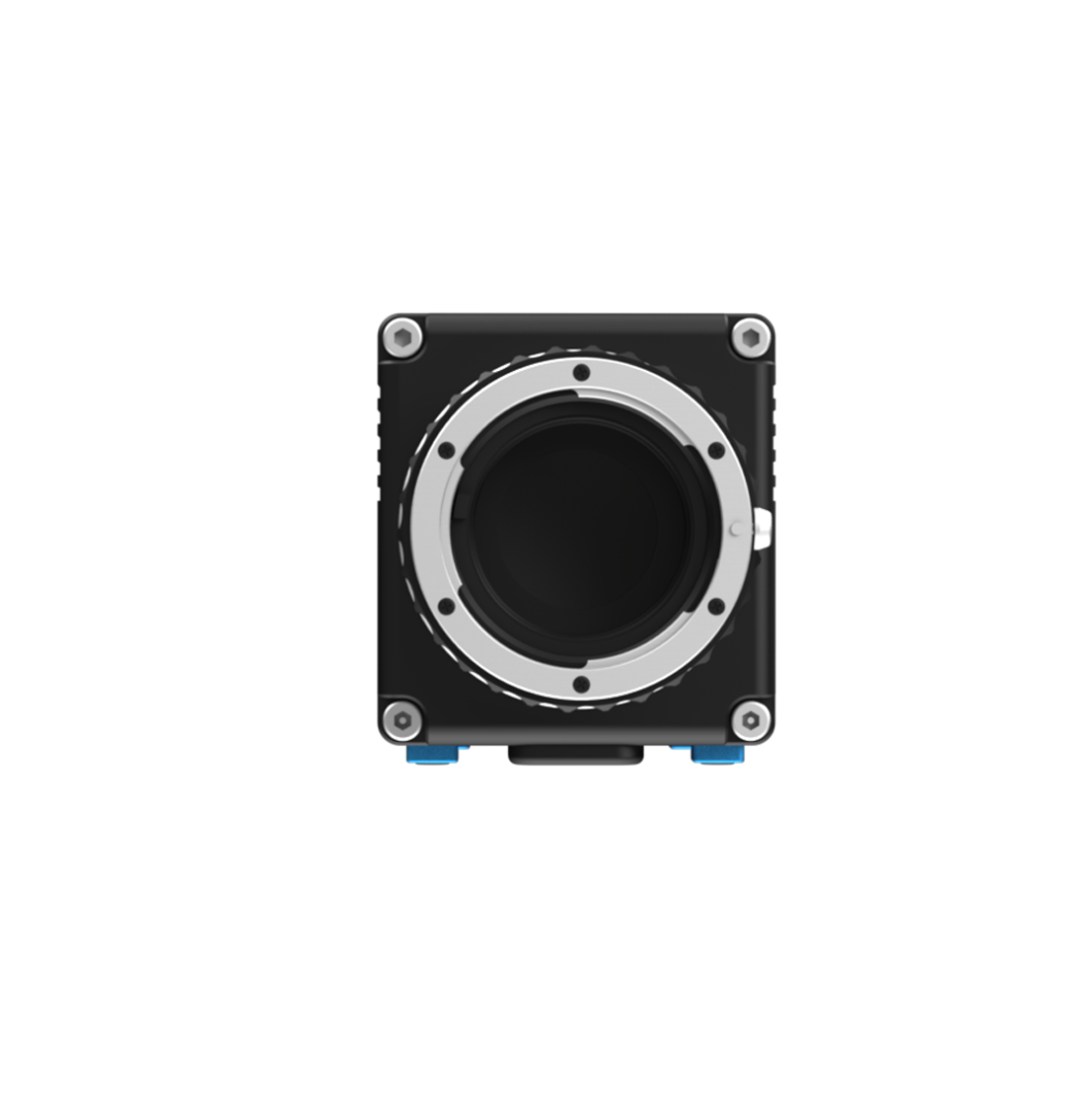
Front View
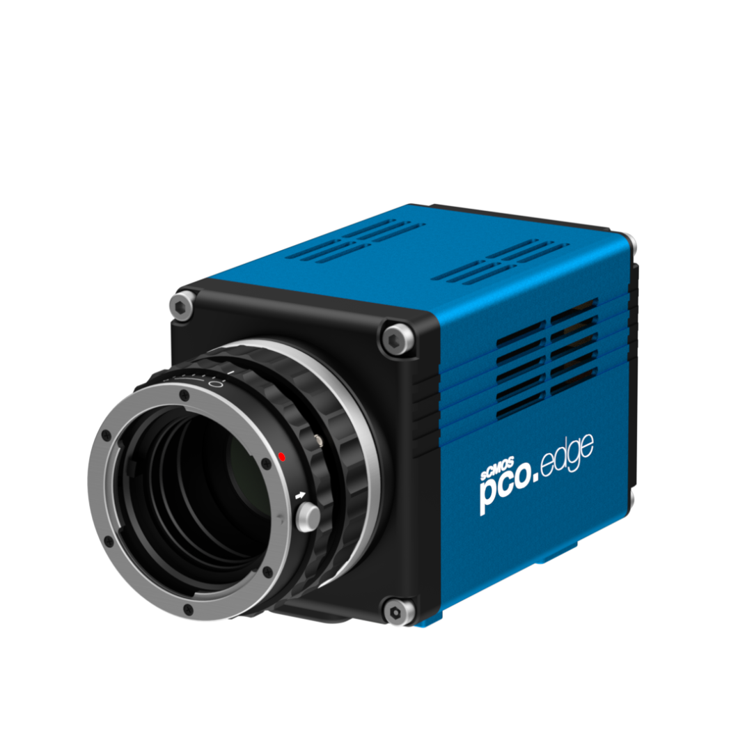
Front View 45 Degree
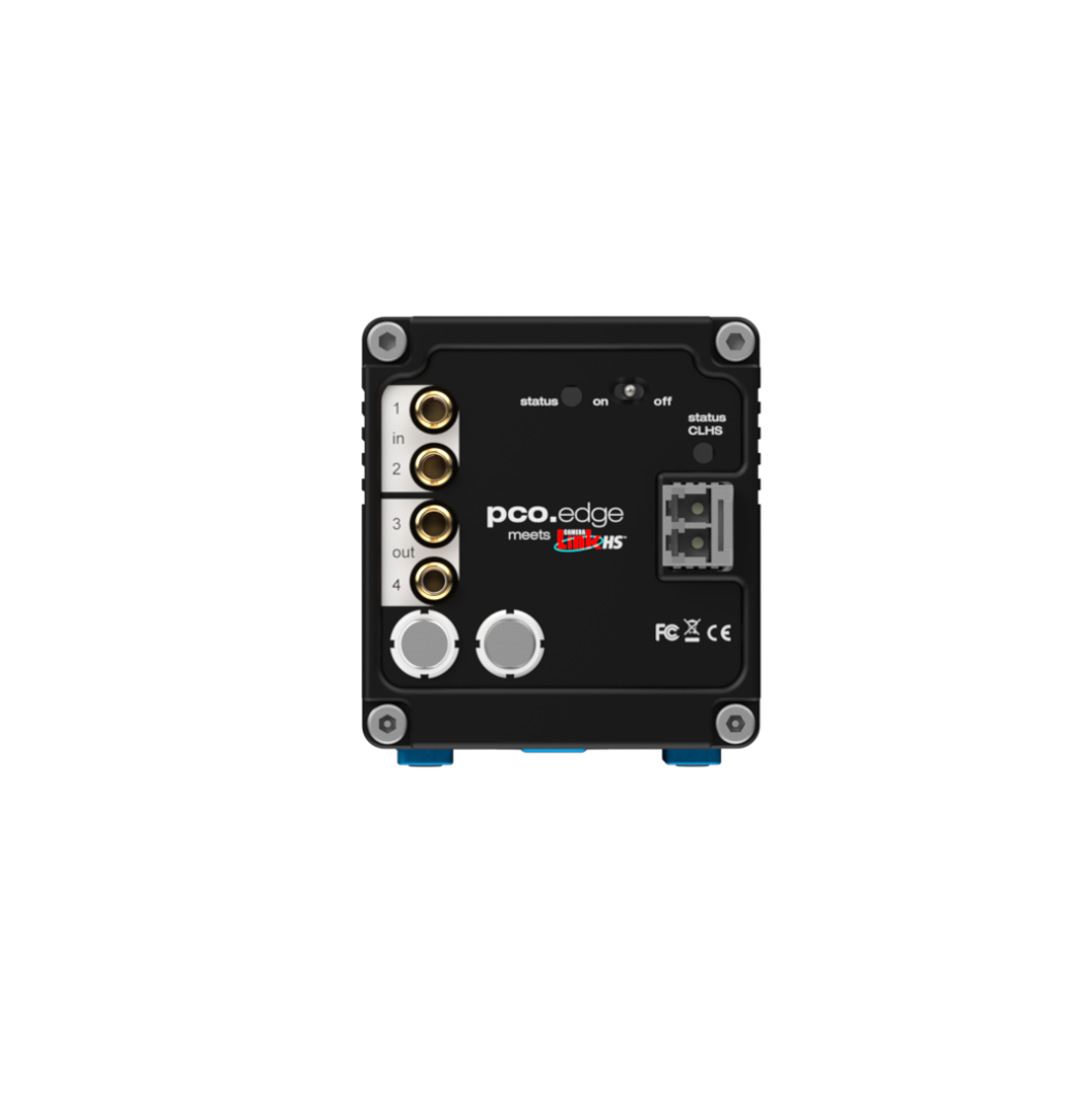
Rear View
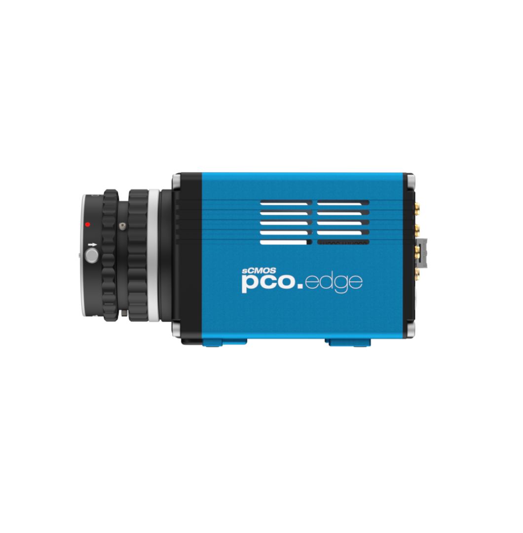
Side View
| Interface | CLHS FOL |
| Sensor technology | sCMOS |
| Color type | monochrome |
| Resolution | 2048 x 2048 pixels |
| Sensor diagonal | 18.8 mm |
| Pixel size | 6.5 x 6.5 μm |
| Maximum frame rate @ full resolution | 100 fps |
| Maximum pixel rate | 548 MPixel/s |
| Peak QE | 82 % @ 580 nm |
| Typical read noise1 | 0.8 e- |
| Dark current @ sensor temperature | < 0.6 @ 7 °C e-/pixel/s |
| Maximum dynamic range | 37,500 : 1 |
| Shutter type | Rolling Shutter |
| Sensor cooling2 | air, optional: water |
| Additional options | lens control |
| Dimensions (H x W x L) | 76 x 70 x 122 mm |
- Bright-field microscopy
- Fluorescence microscopy
- Digital pathology
- Single molecule localization microscopy (SMLM) – PALM, STORM, dSTORM, GSDIM
- Lightsheet fluorescence microscopy (LSFM)
- Structured illumination microscopy (SIM)
- Calcium imaging
- Förster resonance energy transfer (FRET)
- Fluorescence recovery after photobleaching (FRAP)
- High-speed bright-field ratio imaging
- High throughput screening | high content screening
- Biochip reading
- Total internal reflection microscopy (TITF)
- Spinning disk confocal microscopy
- 3D metrology
- Ophthalmology
- Photovoltaic inspection
- Industrial quality inspection
- Wafer inspection
- Image intensifier imaging
- Lucky astronomy
- Particle tracking velocimetry (PTV)
Front View
Front View 45 Degree
Rear View
Side View
Subscribe Now!
Floating Web Forms block
Join the PCO Circular newsletter for early access to insights on our high‑performance imaging cameras. Receive the latest application notes, expert webinars, technical articles, educational videos, product innovations, and industry events designed for scientific, microscopy, research, and industrial imaging applications.