PART/ 85108072513
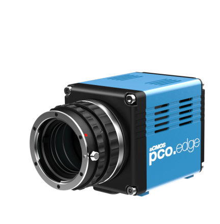
pco.edge 4.2 USB sCMOS Camera
 Powered by Bioz
The pco.edge 4.2 USB scientific camera is equipped with an innovative scientific CMOS (sCMOS) sensor that allows exceptional image quality and precise measurements. With the ability to capture crisp images, the pco.edge 4.2 USB sCMOS camera system is ideal for applications requiring highest quantum efficiency, best 16-bit dynamic range, high frame rates, long exposure times, and extremely low readout noise.
With a very high quantum efficiency (QE), the pco.edge 4.2 USB camera is the ideal choice when high sensitivity is required. The easy-to-connect USB 3.0 interface allows this camera to be used on any device, even notebooks. The sensor is stabilized to 0 °C to reduce the influence of noise and guarantee highest image quality.
| Sensor Technology | sCMOS FSI |
| Resolution (H x V) | 2048 x 2048 |
| Pixel Size (H x V) | 6.5 x 6.5 |
| Sensor Diagonal | 18.8 mm |
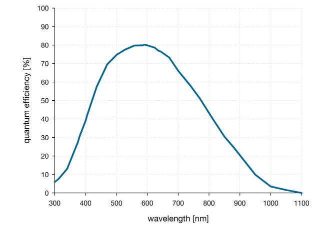
| Peak Quantum Efficiency | 80.0 % |
| Peak Quantum Efficiency Wavelength | 580.0 nm |
| Global Shutter | false |
| Rolling Shutter | true |
| Global Reset | true |
| Operational Modes |
Rolling Shutter with Selectable Readout Direction Global Reset |
| Readout Noise RMS | 1.5 e- |
| Readout Noise Median | 0.8 e- |
| Fullwell Capacity | 30000 e- |
| Dynamic Range | 37500:1 |
| Dynamic Range (log) | 91.0 dB |
| Dark Current | 0.3 e-/s |
| Sensor Temperature (Dark Current Reference) | 0.0 °C |
| Frame rate (Full Sensor) | 40.0 fps |
| Dynamic Range A/D Converter | 16 bit |
| Thermo Electric Cooling | true |
| Forced Air | true |
| Liquid Cooling | true |
| Working Temperature Image Sensor | 0 °C stabilized (up to +27 °C ambient temperature) |
| Data Interface | USB 3.0 |
| Included Lens Mount |
C-Mount F-Mount |
| Width | 70.0 mm |
| Length | 99.5 mm |
| Weight (kg) | 0.75 |
| Height | 76.0 mm |
| Part Number | 85108072513 |
- Bright-field microscopy
- Fluorescence microscopy
- Digital pathology
- Single molecule localization microscopy (SMLM) – PALM, STORM, dSTORM, GSDIM
- Lightsheet fluorescence microscopy (LSFM)
- Structured illumination microscopy (SIM)
- Calcium imaging
- Förster resonance energy transfer (FRET)
- Fluorescence recovery after photobleaching (FRAP)
- High-speed bright-field ratio imaging
- High throughput screening | high content screening
- Biochip reading
- Total internal reflection microscopy (TITF)
- Spinning disk confocal microscopy
- 3D metrology
- Ophthalmology
- Photovoltaic inspection
- Industrial quality inspection
- Wafer inspection
- Image intensifier imaging
- Lucky astronomy
- Particle tracking velocimetry (PTV)
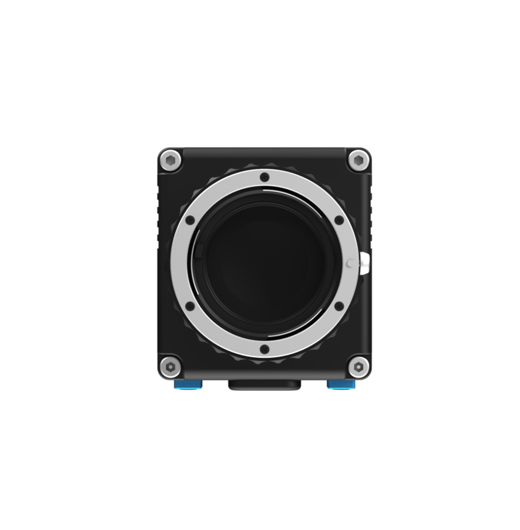
Front View
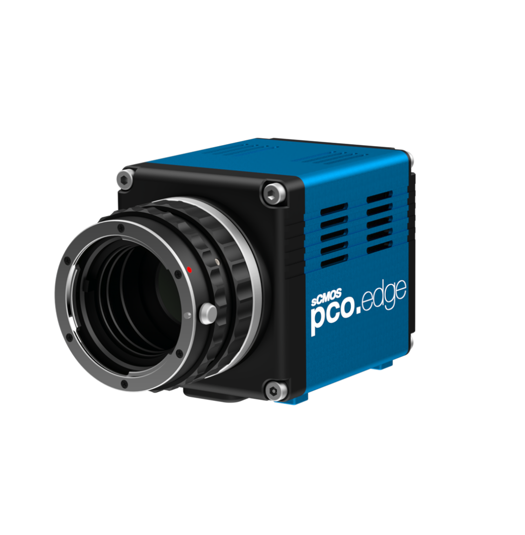
Front View 45 Degree
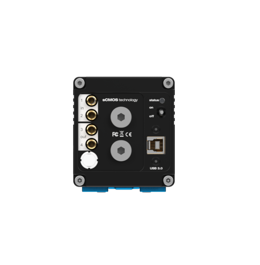
Rear View
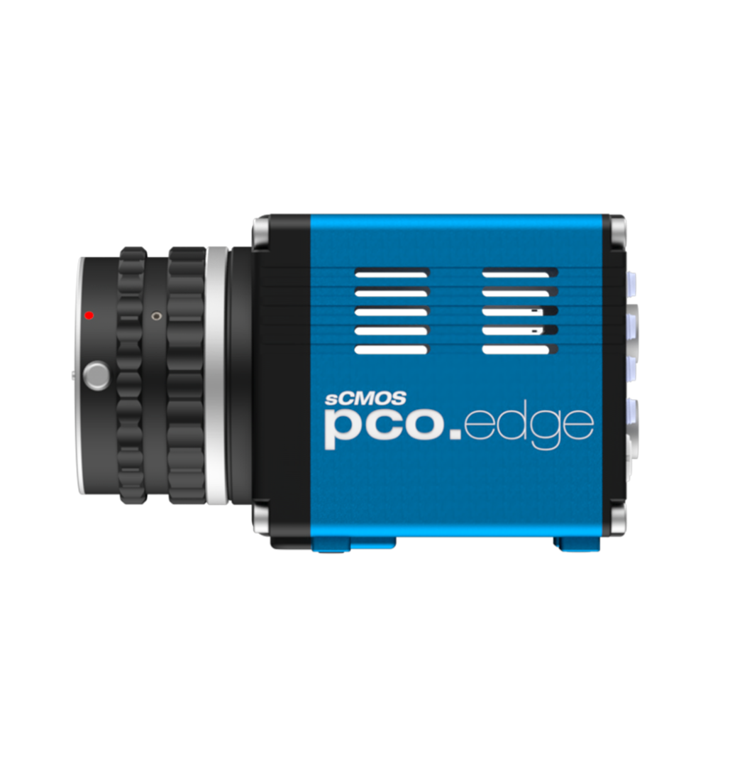
Side View
| Sensor Technology | sCMOS FSI |
| Resolution (H x V) | 2048 x 2048 |
| Pixel Size (H x V) | 6.5 x 6.5 |
| Sensor Diagonal | 18.8 mm |
| Peak Quantum Efficiency | 80.0 % |
| Peak Quantum Efficiency Wavelength | 580.0 nm |
| Global Shutter | false |
| Rolling Shutter | true |
| Global Reset | true |
| Operational Modes |
Rolling Shutter with Selectable Readout Direction Global Reset |
| Readout Noise RMS | 1.5 e- |
| Readout Noise Median | 0.8 e- |
| Fullwell Capacity | 30000 e- |
| Dynamic Range | 37500:1 |
| Dynamic Range (log) | 91.0 dB |
| Dark Current | 0.3 e-/s |
| Sensor Temperature (Dark Current Reference) | 0.0 °C |
| Frame rate (Full Sensor) | 40.0 fps |
| Dynamic Range A/D Converter | 16 bit |
| Thermo Electric Cooling | true |
| Forced Air | true |
| Liquid Cooling | true |
| Working Temperature Image Sensor | 0 °C stabilized (up to +27 °C ambient temperature) |
| Data Interface | USB 3.0 |
| Included Lens Mount |
C-Mount F-Mount |
| Width | 70.0 mm |
| Length | 99.5 mm |
| Weight (kg) | 0.75 |
| Height | 76.0 mm |
| Part Number | 85108072513 |
- Bright-field microscopy
- Fluorescence microscopy
- Digital pathology
- Single molecule localization microscopy (SMLM) – PALM, STORM, dSTORM, GSDIM
- Lightsheet fluorescence microscopy (LSFM)
- Structured illumination microscopy (SIM)
- Calcium imaging
- Förster resonance energy transfer (FRET)
- Fluorescence recovery after photobleaching (FRAP)
- High-speed bright-field ratio imaging
- High throughput screening | high content screening
- Biochip reading
- Total internal reflection microscopy (TITF)
- Spinning disk confocal microscopy
- 3D metrology
- Ophthalmology
- Photovoltaic inspection
- Industrial quality inspection
- Wafer inspection
- Image intensifier imaging
- Lucky astronomy
- Particle tracking velocimetry (PTV)
Front View
Front View 45 Degree
Rear View
Side View
Subscribe Now!
Floating Web Forms block
Join the PCO Circular newsletter for early access to insights on our high‑performance imaging cameras. Receive the latest application notes, expert webinars, technical articles, educational videos, product innovations, and industry events designed for scientific, microscopy, research, and industrial imaging applications.
Floating Web Forms block