
PART/ 85108074001
pco.panda 4.2 sCMOS Camera
Excelitas pco.panda 4.2 is a scientific CMOS camera which features a 16-bit sCMOS sensor. It offers a high resolution of 2048 x 2048 pixels with 6.5 x 6.5 μm pixel size to ensure excellent image quality. Its ultra-compact camera housing and flexible design with the latest sCMOS technology makes the pco.panda 4.2 ideal for use in a wide range of applications.
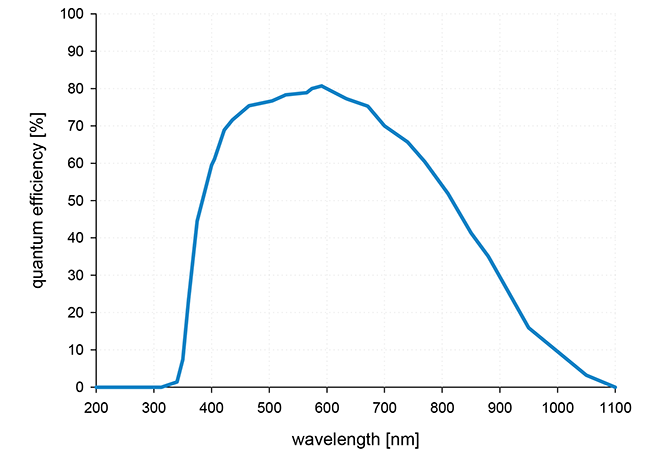
The USB 3.1 connection eliminates the need for an external power supply allowing for faster data transfer and direct power via USB cable. The pco.panda 4.2 has a high quantum efficiency of up to 80 % and low readout noise.
| Interface | USB 3.1 Gen 1 |
| Sensor technology | sCMOS |
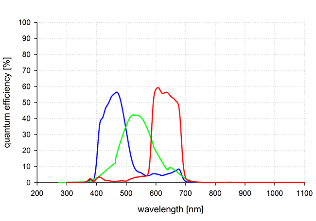
| Color type | monochrome or color |
| Resolution | 2048 x 2048 pixels |
| Sensor diagonal | 18.8 mm |
| Pixel size | 6.5 x 6.5 μm |
| Maximum frame rate | 40 fps |
| Maximum pixel rate | 176 MPixel/s |
| Peak QE | 80 % @ 600 nm 1 |
| Typ. read noise 2 (e-) | 2.1 e - |
| Maximum dynamic range | 21,400 : 1 |
| Shutter type | Rolling Shutter |
| Sensor cooling | passive |
| Additional options 3 | lightsheet scanning mode |
| Dimensions (H x W x L) | 65 x 65 x 66 mm |
- Brightfield microscopy Fluorescence microscopy
- Digital pathology
- Single molecule localization microscopy
- Lightsheet fluorescence microscopy (LSFM)
- Calcium imaging
- FRET
- FRAP
- Structured illumination microscopy (sim)
- High-speed bright field ratio imaging
- High-throughput screening
- High content screening
- Biochip reading
- TIRF microscopy
- Spinning disk confocal microscopy
- Ophthalmology Industrial quality inspection
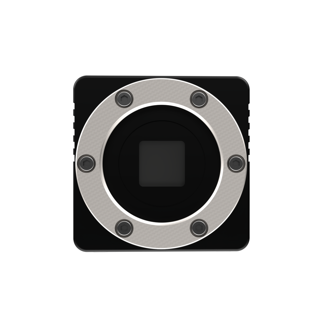
Front View
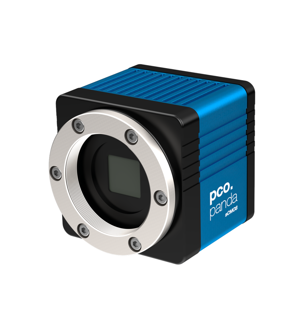
Front View 45 Degree
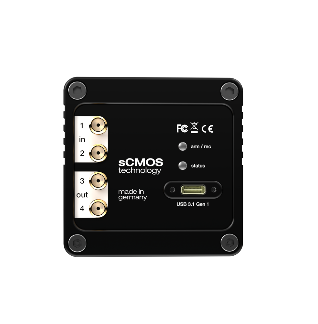
Rear View
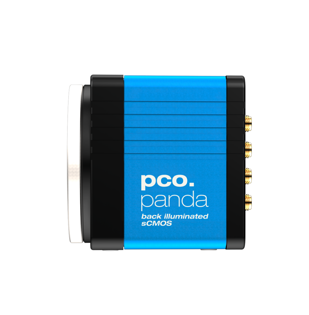
Side View
| Interface | USB 3.1 Gen 1 |
| Sensor technology | sCMOS |
| Color type | monochrome or color |
| Resolution | 2048 x 2048 pixels |
| Sensor diagonal | 18.8 mm |
| Pixel size | 6.5 x 6.5 μm |
| Maximum frame rate | 40 fps |
| Maximum pixel rate | 176 MPixel/s |
| Peak QE | 80 % @ 600 nm 1 |
| Typ. read noise 2 (e-) | 2.1 e - |
| Maximum dynamic range | 21,400 : 1 |
| Shutter type | Rolling Shutter |
| Sensor cooling | passive |
| Additional options 3 | lightsheet scanning mode |
| Dimensions (H x W x L) | 65 x 65 x 66 mm |
- Brightfield microscopy Fluorescence microscopy
- Digital pathology
- Single molecule localization microscopy
- Lightsheet fluorescence microscopy (LSFM)
- Calcium imaging
- FRET
- FRAP
- Structured illumination microscopy (sim)
- High-speed bright field ratio imaging
- High-throughput screening
- High content screening
- Biochip reading
- TIRF microscopy
- Spinning disk confocal microscopy
- Ophthalmology Industrial quality inspection
Front View
Front View 45 Degree
Rear View
Side View
Subscribe Now!
Floating Web Forms block
Join the PCO Circular newsletter for early access to insights on our high‑performance imaging cameras. Receive the latest application notes, expert webinars, technical articles, educational videos, product innovations, and industry events designed for scientific, microscopy, research, and industrial imaging applications.
 Powered by Bioz